Dry Eye Examination
Simplified
From capture to review and follow-up.


Powered by multimodal imaging,
AI-assisted visualization,
and connected workflows.


Integrated Imaging
Enable Multimodal Ocular Imaging
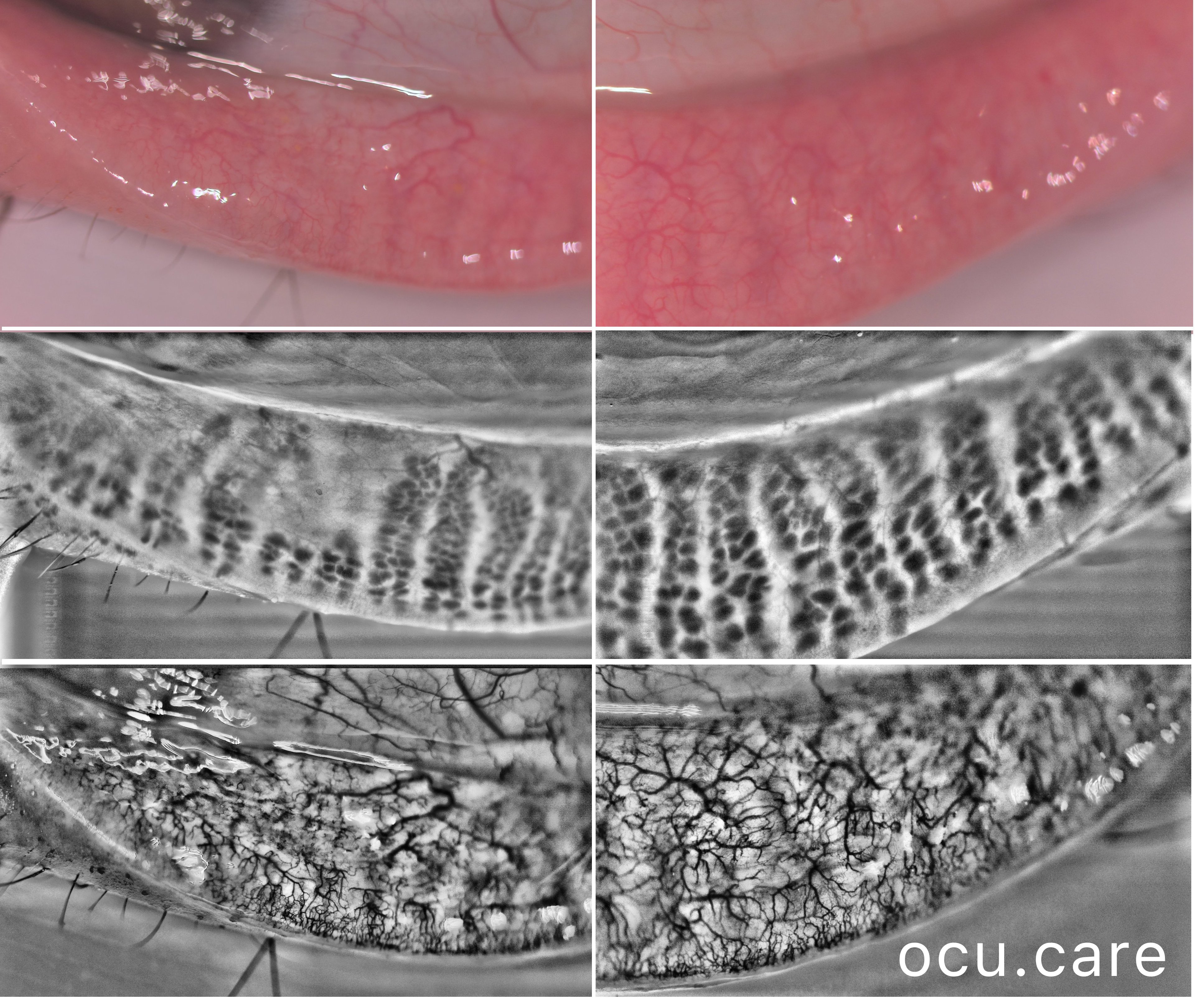
Capture and visualize meibomian gland structure, blink dynamics, tear film appearance, fluorescein imaging, and patient-reported symptoms using a single, compact device designed to integrate seamlessly into existing clinical setups.
AI-Enhanced Visualization
See More with AI-Assisted
Real-Time Visualization
AI-assisted image processing enhances the visibility of ocular surface features, reducing time required manual image adjustments and allowing clinicians to focus on efficient clinical review. Operating directly on the live video stream, it provides immediate visual feedback that supports faster alignment, more confident image capture, and a smoother workflow.


Consistent Workflow
Bring Consistency to Dry Eye Examination
Guided software workflows support consistent imaging and documentation across patients and care settings, helping standardize how dry eye examinations are performed while keeping clinical interpretation in the hands of the clinician.
Data & Connectivity
Share, Store, and Stay Operational
A secure, FHIR- and DICOM-compliant cloud portal with optional on-premise support ensures access to imaging and documentation across practices, even during connectivity outages.

Why MEA?
Efficient & Consistent
Guided workflows streamline imaging and documentation, supporting efficient exam preparation and clinician review.
Unified Exam Report
Clinician-entered notes, imaging, AI-enhanced visualizations, and patient questionnaire responses (e.g. OSDI-6) combined in a single export.
MEA Variants


MEA Capabilities Overview
Powered by proprietary patent-pending technology.
| MEA SL | MEA | |
|---|---|---|
| Supported Modalities | ||
| Conjunctival redness | ✓ | ✓ |
| Blepharitis | ✓ | – |
| Meibomian gland imaging | ✓ | ✓ |
| Ocular surface staining (fluorescein / lissamine green) | ✓ | ✓ |
| User-defined photos | ✓ | ✓ |
| Lipid layer thickness | £* | – |
| Non-invasive tear break-up time (NIBUT) | £* | – |
| Blink rate & quality | £ | £ |
| Tear meniscus height | £* | £* |
| Imaging & Optics | ||
| Blue light illumination | ✓ | ✓ |
| Yellow barrier filter | ✓ | ✓ |
| Workflow & Connectivity | ||
| Slit lamp imaging mode | ✓ | ✓ |
| Handheld imaging mode | – | ✓ |
| Capture → review → follow-up workflow | ✓ | ✓ |
| Automated report generation | ✓ | ✓ |
| Integrated OSDI-6 dry eye questionnaire | ✓ | ✓ |
| Integrated Nathan Efron grading scale | ✓ | ✓ |
| Integrated Heiko Pult meibography grading scale | ✓ | ✓ |
| Cloud portal integration | £ | £ |
| Local DICOM integration | £ | £ |
✓ Included £ Optional module/service * Coming soon – Not available